Look Beyond Hysterectomy and Discover UFE
A highly effective, minimally invasive procedure, UFE typically takes less than an hour to perform. Clinically proven to reduce the major symptoms of uterine fibroids, UFE has become one of the most successful alternatives to hysterectomy procedures.
Interested in learning all about the procedure? Click the play button below to watch the video, then ask your doctor if UFE is the right treatment for you.
The UFE Procedure
Uterine fibroid embolization, also known as uterine artery embolization, is clinically proven to reduce the major symptoms of fibroids, including pelvic pain and pressure, excessive and prolonged bleeding, and frequent urination. It is often performed as an outpatient procedure and the procedure itself lasts about an hour. Compared to a larger surgical incision, UFE is performed through a tiny pinhole like puncture in the upper thigh to provide access to the arteries feeding the fibroids.
UFE is performed by an Interventional Radiologist (IR). Interventional radiologists are medical doctors with additional six or seven years of specialized training after medical school requiring sub-specialized fellowship training after a diagnostic radiology residency. They are certified by the American Board of Radiology. The concept behind interventional radiology is to treat patients using the least invasive techniques currently available to minimize risk to patients and improve health outcomes. Utilizing contemporary imaging techniques, interventional radiologists can effectively treat various complex medical conditions through a pinhole incision with unprecedented precision. This results in reduced risk, less pain, and shorter recovery times. Many of the conditions treated with these techniques involve the vascular system and because of this focus, Interventional Radiologists are considered Vascular Specialists.
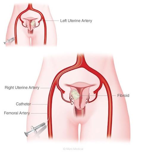
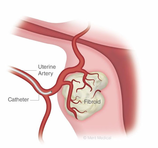
Uterine fibroid embolization, begins with a tiny pinhole like puncture in the groin area. This puncture provides the Interventional Radiologist (IR) with access to arteries that feed the fibroids. Using specialized X-ray equipment, the IR passes a catheter (small tube) into the pinhole like puncture to the uterine artery, and guides it near the location of the fibroid tumor.
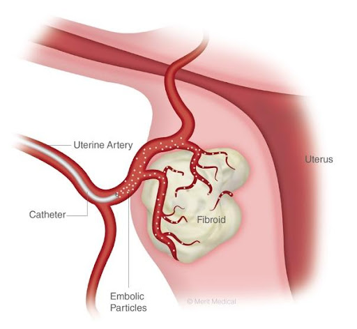
When the IR has reached the location of the fibroids, embolic material (small spheres) are injected through the catheter and into the blood vessels feeding the fibroid, depriving it of oxygenated blood.
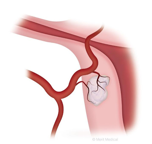
The oxygen deprivation results in fibroids shrinking. The embolic material remains permanently in the blood vessels at the fibroid site. The catheter is then moved to the other side of the uterus. Once the IR has completed embolization of the uterine artery on both sides, the catheter is gently removed.
The entire UFE treatment typically lasts less than one hour, and is typically performed as an outpatient procedure.
Proven Clinical History
UFE was first performed in 1994 and has an extensive clinical history. Hundreds of thousands of women worldwide have been treated with the procedure.
Clinical studies show that UFE provides substantial improvement in major symptoms, including pelvic pain and discomfort, heavy menstrual bleeding, and urinary problems. Three-year data from the Fibroid Registry for Outcomes Data (FIBROID) indicated that UFE offers sustained improvement in quality of life and symptom relief—90 percent of the women participating avoided a hysterectomy, of which 85 percent had substantial improvements in symptoms and quality of life.
Imagine a life free of fibroid symptoms!
© Merit Medical, Used With Permission.