For many women experiencing pelvic discomfort or irregular menstrual cycles, a fibroid ultrasound is often the first step in identifying uterine fibroids. At 1Fibroid, serving Manhattan, Queens, and Rego Park, NY, patients can gain clarity about their condition through this non-invasive imaging technique. Understanding what to expect during the procedure can help alleviate anxiety and ensure patients feel informed and prepared. A fibroid ultrasound is a painless diagnostic tool that provides detailed images of the uterus, helping doctors detect the size, number, and location of fibroids.
Ultrasounds use high-frequency sound waves to create images of internal organs, making them safe and widely used in gynecological care. The procedure is typically quick, usually lasting between 20 and 40 minutes, and does not involve radiation. Being well-informed about the process—from preparation to post-procedure results—can help women feel more comfortable and empowered during their visit.
Preparing for a Fibroid Ultrasound
Preparation for a fibroid ultrasound is straightforward but important for obtaining accurate images. Patients may be asked to drink water and have a full bladder before the exam. A full bladder helps move the intestines out of the way and provides a clearer view of the uterus. Some patients may also receive instructions to avoid certain foods or beverages if a transabdominal approach is used.
It’s also important for patients to wear comfortable, loose-fitting clothing, as this makes it easier to access the abdominal area for the ultrasound probe. Women should be prepared to provide a detailed medical history, including information about menstrual cycles, previous surgeries, and any symptoms they may be experiencing. Having this information readily available helps the provider interpret ultrasound results accurately and tailor recommendations based on the patient’s unique health profile.
The Ultrasound Procedure
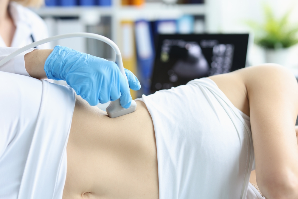
The procedure itself can be performed in one of two ways: transabdominally or transvaginally. During a transabdominal ultrasound, a gel is applied to the lower abdomen to help sound waves pass smoothly, and the provider moves a handheld device called a transducer across the skin to capture images of the uterus. Transvaginal ultrasounds involve inserting a smaller probe into the vagina, offering a closer view of the uterus and fibroids.
During the exam, patients typically lie on their back on an exam table. While the procedure is non-invasive, some may feel mild pressure or discomfort, especially if a full bladder is required. Providers take multiple images from different angles to ensure a comprehensive view. The process is generally well-tolerated, and patients can usually resume normal activities immediately afterward.
Understanding Ultrasound Results
After the ultrasound, the images are analyzed by a radiologist or the provider to determine the presence, size, and location of fibroids. Results may also indicate other conditions such as cysts or uterine abnormalities. The findings are usually discussed in a follow-up appointment, where patients can ask questions and receive guidance on next steps or treatment options.
It is important to remember that ultrasound provides valuable information but is just one part of a larger diagnostic process. Other tests or evaluations may be recommended based on the ultrasound findings. Having a clear understanding of the results can help patients make informed decisions about their reproductive health and overall well-being.
Tips for a Comfortable Experience
- Wear loose-fitting clothing for easy access to the abdominal area
- Drink plenty of water if instructed to have a full bladder
- Communicate any discomfort or concerns during the procedure
- Bring a list of current medications and medical history for accuracy
- Ask questions about the process or results to feel informed and empowered
Following these tips can help ensure a smooth and comfortable experience during the fibroid ultrasound. Patients often find that knowing what to expect reduces anxiety and improves the overall experience.
Conclusion
A fibroid ultrasound is a safe and informative procedure that provides critical insight into uterine health. Women in Manhattan, Queens, and Rego Park, NY, can benefit from understanding each step of the process, from preparation to receiving results. Being informed helps reduce anxiety, ensures accurate imaging, and empowers patients to take an active role in their health. At 1Fibroid, the goal is to provide patients with clear information and support throughout their diagnostic journey.
Resources
- Stewart, E. A. (2001). Uterine Fibroids. New England Journal of Medicine.
- Buttram, V. C., & Reiter, R. C. (1981). Uterine Leiomyomata: Etiology, Symptomatology, and Management. Fertility and Sterility.
- Parker, W. H. (2007). Etiology, Symptomatology, and Diagnosis of Uterine Fibroids. Obstetrics & Gynecology.